| Issue |
Knowl. Manag. Aquat. Ecosyst.
Number 417, 2016
Topical issue on Crayfish
|
|
|---|---|---|
| Article Number | 32 | |
| Number of page(s) | 5 | |
| DOI | https://doi.org/10.1051/kmae/2016019 | |
| Published online | 31 August 2016 | |
Short communication
Fine-tuning of a COI PCR-RFLP assay for fast genetic characterization of Spanish white-clawed crayfish
Mise au point d’un essai COI PCR-RFLP pour une caractérisation génétique rapide de l’écrevisse à pattes blanches espagnole
Departamento de Genética, Facultad de Ciencias
Biológicas, Universidad Complutense, C/José Antonio Novais 12, Edificio B, Planta 2,
28040
Madrid,
Spain
⋆ Corresponding author:
This email address is being protected from spambots. You need JavaScript enabled to view it.
Received:
19
April
2016
Accepted:
21
June
2016
Abstract
The white-clawed crayfish is endemic to western and southern Europe and its population has dramatically decreased in the last few decades. The latest reports on this species have shown that the genetic variability of Spanish populations displays a clear geographic pattern, with two main genetic groups across its range of distribution. To ensure the preservation of this endangered species, specimens for restocking purposes should be chosen, ideally, accordingly to their genetic characteristics. Here, we propose a PCR-RFLP assay developed for this purpose. Digestion of a fragment of the mitochondrial cytochrome oxidase subunit I gene – with two restriction enzymes, HpaI and CviAII - differentiates the most common haplotypes identified in Spain. This technique is a useful, low-cost and rapid method to facilitate the genetic characterization of Spanish white-clawed crayfish and, in doing so, to incorporate genetic information into conservation plans for this species.
Résumé
L’écrevisse à pattes blanches est endémique de l’Europe occidentale et méridionale et ses populations ont considérablement diminué au cours des dernières décennies. Les derniers rapports sur cette espèce ont montré que la variabilité génétique des populations espagnoles affiche une répartition géographique claire avec deux principaux groupes génétiques dans son aire de distribution. Pour assurer la conservation de cette espèce en voie de disparition, les échantillons à des fins de repeuplement doivent être choisis, idéalement, en fonction de leurs caractéristiques génétiques. Nous proposons ici une analyse PCR-RFLP développée à cet effet. La digestion d’un fragment du gène de la sous-unité I de cytochrome oxydase mitochondriale - avec deux enzymes de restriction HpaI et CviAII - différencie les haplotypes les plus communs identifiés en Espagne. Cette technique est une méthode, à faible coût, utile et rapide pour faciliter la caractérisation génétique de l’écrevisse à pattes blanches espagnole et, ce faisant, pour incorporer l’information génétique dans les plans de conservation pour cette espèce.
Key words: Spanish white-clawed crayfish / Cytochrome oxidase subunit I (COI) / PCR–RFLP / Restriction enzymes / genetic groups
Mots clés : écrevisse à pattes blanches espagnole / Cytochrome oxydase sous-unité I (COI) / PCR-RFLP / enzyme de restriction / groupes génétique
© B. Matallanas et al., published by EDP Sciences, 2016
 This is an Open Access article distributed under the terms of the Creative
Commons Attribution License CC-BY-ND (http://creativecommons.org/licenses/by-nd/4.0/), which permits
unrestricted use, distribution, and reproduction in any medium, provided the original
work is properly cited. If you remix, transform, or build upon the material, you may not
distribute the modified material.
This is an Open Access article distributed under the terms of the Creative
Commons Attribution License CC-BY-ND (http://creativecommons.org/licenses/by-nd/4.0/), which permits
unrestricted use, distribution, and reproduction in any medium, provided the original
work is properly cited. If you remix, transform, or build upon the material, you may not
distribute the modified material.
1 Introduction
Preserving threatened species is a challenge nowadays. Accelerating climate change and the increasing effects of human activities have an enormous impact on many regions, having particularly affected the freshwater ecosystems of the Mediterranean Basin.
Crayfish are considered a keystone species because they play an important role in freshwater ecosystems (Geiger et al., 2005), simultaneously acting as both prey and predator. The white-clawed crayfish (formally described as Austropotamobius pallipes sensu lato), endemic to Western Europe, has had a progressive and drastic decrease in its population all over its distribution range, and especially in Spain (Alonso et al., 2000). This decrease has been due to many factors such as habitat destruction and introduction of alien species. Consequently, the species benefits from protection under regional, national and international legislation.
Given its ecological relevance, current protection and conservation measures should be maintained, but these programs are mainly based on reintroduction and translocation of specimens. Notwithstanding, genetic information must be included in effective long-term conservation planning, if efforts are to be effective.
DNA methods based on polymerase chain reaction (PCR) amplification have been successfully used to characterize genetic variability in the white-clawed crayfish (Amouret et al., 2015; Beroiz et al., 2008; Gouin et al., 2006; among many others). PCR-restriction fragment length polymorphism (PCR-RFLP), one of these methods, consists of the amplification of a DNA fragment, followed by restriction enzyme treatment and electrophoretic separation. Specific band profiles allow detecting variation at the DNA level without costly DNA sequencing. PCR-RFLP analysis of mitochondrial DNA (mtDNA) has been used to assess genetic diversity in several crustacean species, such as lobsters (García-Rodríguez et al., 2006), mitten crabs (Cho et al., 2014), shrimps (Bouchon et al., 1994) and crayfish (Soroka, 2008).
The latest reports on Spanish white-clawed crayfish (Matallanas et al., 2016) have identified four common mitochondrial haplotypes and an evident genetic structure of the populations, with two clearly distinguishable genetic groups, ‘Northern’ and ‘Central’. To ensure the preservation of these genetic groups, specimens for restocking purposes should be chosen, ideally, accordingly to their genetic characteristics based on diagnostic genetic markers.
In this study, we present a low-cost, rapid and convenient PCR-RFLP method for genotyping Spanish white-clawed crayfish and identifying the genetic group to which they belong.
Populations of A. pallipes analyzed in the present work. CAN: Cantabrian Basin, MED: Mediterranean Basin, ATL: Atlantic Basin.
2 Material and methods
One hundred and sixty specimens (from sixteen populations) from a previous survey (Matallanas et al., 2016) were selected for this PCR-RFLP assay (Table 1).
Genomic DNA was extracted from 20−50 mg of claw muscle or pereopod tissues using the DNeasy Blood and Tissue Kit from Qiagen (Valencia, CA, USA). The mtDNA COI fragment was amplified in a final volume of 50 ¯L with25 ng of total DNA, 1×reaction buffer, 2 mM MgSO4, 200 ¯M of each dNTP, 15 ¯g of BSA, 1 ¯M of each primer and 1 U of Vent DNA polymerase (New England BioLabs, Ipswich, MA, USA). The primers used were C/N 2769 (Gopurenko et al., 1999) and LCO 1490 (Folmer et al., 1994). The PCR program included an initial denaturation step of 94 °C for 5 min followed by 44 cycles of 94 °C for 45 s, 53 °C for 1 min and 72 °C for 1 min 30 s, plus a final extension step of 72 °C for 10 min.
After sequencing at the Genomics Unit of the Universidad Complutense de Madrid, the COI fragments were aligned using the CLUSTAL W algorithm (Thompson et al., 1997) and edited with BioEdit v.7.0.9.0 software (Hall, 1999). To identify the number of different haplotypes and their frequencies, the DnaSP v5.10 program was used (Librado and Rozas 2009).
In order to find restriction sites in the sequences, restriction maps of COI haplotypes were screened with over two hundred different enzymes using the SeqBuilder® v.7 program (DNAStar, Madison, WI, USA). Several suitable enzymes were tested, and finally two, HpaI and CviAII, were selected for the RFLP assay.
The HpaI restriction reactions were performed in a final volume of 25 ¯L with 5 ¯L of PCR product, 1x reaction buffer and 6 U of HpaI (New England BioLabs, Ipswich, MA, USA), incubated for 24 h at 37 °C. The CviAII reactions were carried out in a final volume of 25 ¯L with 5 ¯L of PCR product, 1x reaction buffer, 1.6 mg·mL-1 of BSA (Bovine Serum Albumin, Fraction V, Roche Applied Science, Mannheim, Germany) and 16 U of CviAII (New England BioLabs, Ipswich, MA, USA), incubated at 25 °C for 24 h. The reactions were stopped by adding 5 ¯L of loading buffer. The whole reaction volume was then loaded on 3% agarose gel, stained with ethidium bromide (0.5 ¯L·mL-1) and visualized in an E-BOX-VX2/20M gel documentation system (Vilber Lourmet, Torcy, France).
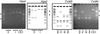 |
Fig. 1 Observed and expected restriction patterns of the COI PCR products of A. pallipes samples in Spanish populations with HpaI and CviAII. Lines 1 and 7 – in each gel – and lines 1 and 6 – in each scheme – show the GeneRulerTM 100 bp DNA Ladder (Thermo Scientific). Asterisks indicate the 500 bp and 1000 bp bands. Patterns A, B, C and D are detailed in Table 1. Ctrl: Restriction control. |
3 Results
The primer pair of the mitochondrial COI gene yielded amplicons of around 1200 bp in length. The HpaI restriction enzyme recognized zero or one restriction site(s) within the COI amplicons, resulting in two easily distinguishable patterns (I and II). Endonuclease CviAII had four or five sites in the COI fragment studied, allowing the detection of three different restriction fragment patterns (α, β and γ) (Table 2 and Figure 1).
Expected fragment sizes (in base pairs, bp) from restriction of COI PCR products with HpaI and CviAII according to the 1184 pb fragment of the COI haplotypes detected in the white-clawed crayfish specimens.
As a whole, the complete COI PCR-RFLP assay yielded four different restriction patterns (A, B, C and D) (Table 2). According to these restriction patterns, crayfish can be assigned to a genetic group. This PCR-RFLP assay roughly discriminates the four most common haplotypes previously found in Spain, given that they account for nearly 96% of the crayfish analyzed. The remaining seven exclusive haplotypes detected could not be identified. In these private haplotypes, the endonucleases do not have restriction sites at the positions where these SNPs occur. Nevertheless, these population-specific haplotypes were present at very low frequencies. In such cases, even though the COI haplotype cannot be detected exactly, the crayfish can be assigned to a genetic group.
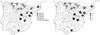 |
Fig. 2 Distribution of the most common COI haplotypes and restriction patterns found in the white-clawed crayfish populations analyzed. Pie graphs represent the relative proportion of COI haplotypes (A) and restriction patterns (B) for each population sampled. |
4 Discussion
For the last few years, the genetic variability of Spanish white-clawed crayfish has been assessed with mitochondrial and nuclear markers by sampling a significant number of populations throughout its distribution range in the country (Beroiz et al., 2008; Matallanas et al., 2013, 2016; Pedraza-Lara et al., 2010).
The COI PCR-RFLP assay presented here enables the detection of the most common haplotypes in Spanish crayfish previously identified by sequencing, in a fast and cost-effective way. The restriction patterns C and D identify the EF485041 and FJ897842 COI haplotypes detected in populations of white-clawed crayfish from Northern Spain. Patterns A and B correspond to the FJ897841 and FJ897840 COI haplotypes, characteristic of the rest of the country (Figure 2) (accession numbers EF485041 and FJ897840-42 correspond to haplotypes deposited in the GenBank database).
Previous reports showed the suitability of COI PCR-RFLP assays to discriminate crayfish species (Astacus astacus, A. leptodactylus, Pacifastacus leniusculus and Orconectes limosus). However, after testing up to six restriction endonucleases, the technique, according to the author, “did not detect any specimen variability within the species” (Soroka, 2008). Particularly, the mitochondrial genome of the white-clawed crayfish (A. pallipes species complex) was analyzed with RFLP with a variable number of enzymes (six to eleven) (Grandjean et al., 1996, 1997, 2000a, 2000b, 2001). However, the results of these experiments did not show that Spanish crayfish had inter- or intrapopulation genetic variation (Grandjean et al., 1997, 2001).
Conversely, the two restriction enzymes selected for this assay – after screening for over two hundred different enzymes – can reveal the genetic variability of the Spanish populations of white-clawed crayfish. Therefore, this PCR-RFLP assay significantly reduces the cost associated with the analysis. Furthermore, crayfish can be assigned to one of the two genetic groups described in Spain (Matallanas et al., 2016; Figure 2).
In summary, the results demonstrate that this COI PCR-RFLP assay is a very advantageous and useful technique for analyzing Spanish populations of white-clawed crayfish. Previous studies showed that Spanish white-clawed crayfish should constitute an independent ESU (Evolutionary Significant Unit, Moritz, 1994). This ESU exhibits two management units (MUs) (Moritz, 2002), named the ‘Northern’ and ‘Central’ groups according to their geographical distribution in Spain. Hence, based on this geographical pattern of mitochondrial variation and taking into account the genetic information available, the introduction of crayfish specimens to a host population should be made with caution.
The assay presented here is an easy, cheap and fast tool for genetic characterization of crayfish. This COI PCR-RFLP assay can contribute to devising conservation program strategies that can efficiently maintain, and even recover in some geographic areas, the genetic variability of this endangered species.
Acknowledgments
This work was funded by the Project MCYT CGL2005-05727/BOS and Convenio MMA-UCM 415-2634. The authors are very grateful to Alonso F., Bassols E., Beroiz B., Bertocchi S., Brusconi S., Cano M.C., Casanova J.M., Castién E., Diéguez-Uribeondo J. , Gil J.M., Juncal J.A., Lamora J., Lasheras I., Lombart A., Martín M.L., Múzquiz J.L., Palacios B., Pinedo J., Sancho V., Temiño C. and Vidal M.I. for providing crayfish samples for this study.
References
- Alonso F., Temiño C. and Diéguez-Uribeondo J., 2000. Status of the white-clawed crayfish Austropotamobius pallipes (Lereboullet 1858), in Spain distribution and legislation. Bull. Fr. Pêche Piscic., 356, 31–54. [CrossRef] [EDP Sciences] [Google Scholar]
- Amouret J., Bertocchi S., Brusconi S., Fondi M., Gherardi F., Grandjean F., Chessa L.A., Tricarico E. and Souty-Grosset C., 2015. The first record of translocated white-clawed crayfish from the Austropotamobius pallipes complex in Sardinia (Italy). J. Limnol., 74, 491–500. [Google Scholar]
- Avise J.C., 2000. Phylogeography: The History and Formation of Species. Harvard University Press, Massachusetts, USA. [Google Scholar]
- Beroiz B., Callejas C., Alonso F., and Ochando, M.D., 2008. Genetic structure of Spanish white-clawed crayfish (Austropotamobius pallipes) populations as determined by RAPD analysis: reasons for optimism. Aquat. Conserv., 18, 190–201. [CrossRef] [Google Scholar]
- Bouchon D., Souty-Grosset C. and Raimond R., 1994. Mitochondrial DNA variation and markers of species identity in two penaeid shrimp species. Penaeus monodon Fabricius and P. japonicus Bate. Aquaculture, 127, 131–144. [CrossRef] [Google Scholar]
- Cho Y.A., Kim E.M., Kim M.J., Kang J.H., Dong C.M., An H.S., An C.M., Park M.A. and Park J.Y., 2014. A rapid and simple method for distinguishing two mitten crabs (Eriocheir sinensis and Eriocheir japonica) in Korea using PCR-RFLP and PCR. Food Control, 36, 20–23. [CrossRef] [Google Scholar]
- Folmer O., Black M., Hoeh W., Lutz R. and Vrijenhoek R., 1994. DNA primers for amplification of mitochondrial cytochrome c oxidase subunit I from diverse metazoan invertebrates. Mol. Mar Biol. Biotechnol., 3, 294–299. [Google Scholar]
- García-Rodríguez F.J. and Perez-Enriquez R., 2006. Genetic differentiation of the California spiny lobster Panulirus interruptus (Randall, 1840) along the west coast of the Baja California Peninsula, Mexico. Mar. Biol., 148, 621–629. [CrossRef] [Google Scholar]
- Geiger W., Alcorlo P., Baltanas A. and Montes C., 2005. Impact of an introduced crustacean on the trophic webs of Mediterranean wetlands. Biol. Invasions, 7, 49–73. [CrossRef] [Google Scholar]
- Gopurenko D., Hughes J.M. and Keenan C.P., 1999. Mitochondrial DNA evidence for rapid colonisation of the Indo-West Pacific by the mud crab Scylla serrata. Mar. Biol., 134, 227–233. [CrossRef] [Google Scholar]
- Gouin N., Grandjean F., Pain S., Souty-Grosset C. and Reynolds J., 2003. Origin and colonization history of the white-clawed crayfish, Austropotamobius pallipes, in Ireland. Heredity, 91, 70–77. [CrossRef] [PubMed] [Google Scholar]
- Gouin N., Grandjean F. and Souty-Grosset C., 2006. Population genetic structure of the endangered crayfish Austropotamobius pallipes in France based on microsatellite variation: biogeographical inferences and conservation implications. Freshw. Biol., 51, 1369–1387. [CrossRef] [Google Scholar]
- Grandjean F. and Souty-Grosset C. 1996. Isolation and characterisation of mitochondrial DNA from the endangered white-clawed crayfish Austropotamobius pallipes, Lereboullet, 1858. Bull. Fr. Pêche Piscic., 343, 175–182. [CrossRef] [EDP Sciences] [Google Scholar]
- Grandjean F. and Souty-Grosset C., 2000a. Genetic and morphological variation in the endangered crayfish species, Austropotamobius pallipes (Lereboullet) (Crustacea, Astacidae) from the Poitou-Charentes region (France). Aquat. Sci., 62, 1–19. [Google Scholar]
- Grandjean F. and Souty-Grosset C., 2000b. Mitochondrial DNA variation and population genetic structure of the white-clawed crayfish, Austropotamobius pallipes pallipes. Conserv. Genet., 1, 309–319. [Google Scholar]
- Grandjean F., Souty-Grosset C., Raimond R. and Holdich D.M., 1997. Geographical variation of mitochondrial DNA between populations of the white-clawed crayfish Austropotamobius pallipes. Freshw. Biol., 37, 493–501. [CrossRef] [Google Scholar]
- Grandjean F., Gouin N., Souty-Grosset C. and Diéguez-Uribeondo J. 2001. Drastic bottlenecks in the endangered crayfish species, Austropotamobius pallipes in Spain with inference to its colonization history. Heredity, 88, 1–8. [Google Scholar]
- Hall T.A., 1999. BioEdit: a user – friendly biological sequence alignment editor and analysis program for Windows 95/98/NT. Nucleic Acids Symp. Ser., 41, 95–98. [Google Scholar]
- Librado P. and Rozas J., 2009. DnaSP v5: a software for comprehensive analysis of DNA polymorphism data. Bioinformatics, 25, 1451–1452. [CrossRef] [PubMed] [Google Scholar]
- Matallanas B., Ochando M.D., Alonso F. and Callejas C., 2013. Phylogeography of the white-clawed crayfish (Austropotamobius italicus) in Spain: inferences from microsatellite markers. Mol. Biol. Rep., 40, 5327–5338. [CrossRef] [PubMed] [Google Scholar]
- Matallanas B., Ochando M.D., Alonso F. and Callejas C., 2016. Update of genetic information for the white-clawed crayfish in Spain, with new insights into its population genetics and origin. Org. Divers. Evol. DOI:10.1007/s13127-016-0268-4 [Google Scholar]
- Moritz C., 1994. Defining ‘Evolutionary Significant Units’ for conservation. Trends Ecol. Evol., 9, 373–375. [Google Scholar]
- Moritz C., 2002. Strategies to protect biological diversity and the evolutionary processes that sustain it. Syst. Biol., 51, 238–254. [CrossRef] [PubMed] [Google Scholar]
- Nantón A., Freire R., AriasPérez A., Gaspar M. B. and Méndez J., 2015. Identification of four Donax species by PCR–RFLP analysis of cytochrome c oxidase subunit I (COI). Eur. Food Res. Technol., 240, 1129–1133. [CrossRef] [Google Scholar]
- Pedraza-Lara C., Alda F., Carranza S. and Doadrio I., 2010. Mitochondrial DNA structure of the Iberian populations of the white – clawed crayfish, Austropotamobius italicus italicus (Faxon, 1914). Mol. Phylogenet. Evol. 57, 327–342. [CrossRef] [PubMed] [Google Scholar]
- Thompson J.D., Gibson T.J., Plewniak F., Jeanmougin F. and Higgins D.G., 1997. The Clustal X window interface: flexible strategies for multiple sequence alignment aided by quality analysis tools. Nucleid Acids Res., 24, 4876–4882. [CrossRef] [Google Scholar]
- Soroka M., 2008. Application of mitochondrial DNA in the identification of diverse crayfish species. Pol. J. Nat. Sci., 23, 624–634. [CrossRef] [Google Scholar]
Cite this article as: B. Matallanas, M.D. Ochando, C. Callejas, 2016. Fine-tuning of a COI PCR-RFLP assay for fast genetic characterization of Spanish white-clawed crayfish. Knowl. Manag. Aquat. Ecosyst., 417, 32.
All Tables
Populations of A. pallipes analyzed in the present work. CAN: Cantabrian Basin, MED: Mediterranean Basin, ATL: Atlantic Basin.
Expected fragment sizes (in base pairs, bp) from restriction of COI PCR products with HpaI and CviAII according to the 1184 pb fragment of the COI haplotypes detected in the white-clawed crayfish specimens.
All Figures
|
Fig. 1 Observed and expected restriction patterns of the COI PCR products of A. pallipes samples in Spanish populations with HpaI and CviAII. Lines 1 and 7 – in each gel – and lines 1 and 6 – in each scheme – show the GeneRulerTM 100 bp DNA Ladder (Thermo Scientific). Asterisks indicate the 500 bp and 1000 bp bands. Patterns A, B, C and D are detailed in Table 1. Ctrl: Restriction control. |
| In the text | |
|
Fig. 2 Distribution of the most common COI haplotypes and restriction patterns found in the white-clawed crayfish populations analyzed. Pie graphs represent the relative proportion of COI haplotypes (A) and restriction patterns (B) for each population sampled. |
| In the text | |
Current usage metrics show cumulative count of Article Views (full-text article views including HTML views, PDF and ePub downloads, according to the available data) and Abstracts Views on Vision4Press platform.
Data correspond to usage on the plateform after 2015. The current usage metrics is available 48-96 hours after online publication and is updated daily on week days.
Initial download of the metrics may take a while.